Nagoya University lab method reads microRNA and protein in single vesicles to locate inflammation
Nagoya University researchers read membrane proteins and microRNAs in intact extracellular vesicles. In lab and patient samples, the method linked inflammation to its tissue of origin.
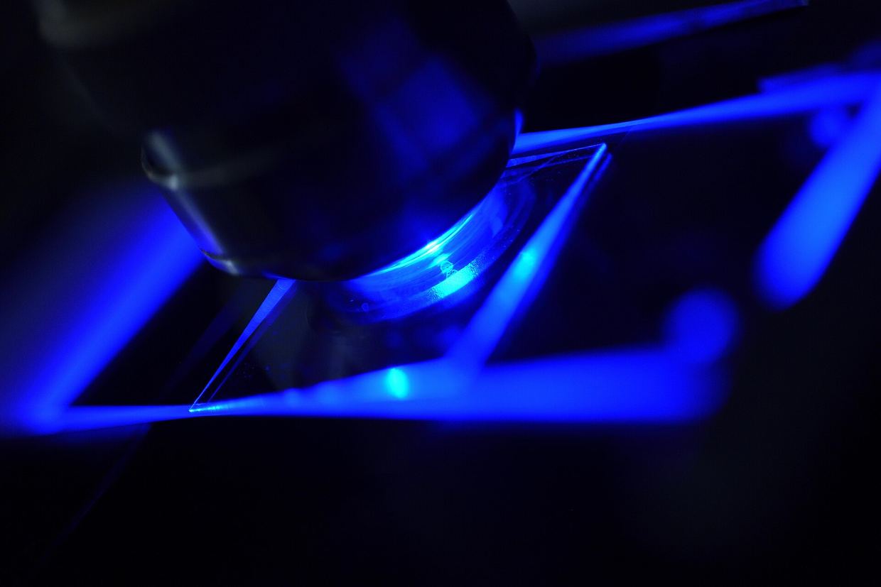
Researchers at Nagoya University in Japan have developed a method that reads membrane proteins and microRNAs in individual extracellular vesicles (EVs) from blood and urine without breaking the vesicles open. The work was published in Cell Reports Physical Science on 17 October 2026 doi.org
Cell Reports Physical Sciencedoi.orgOpen source ↗. The aim is to show not only that chronic inflammation is present, but also in which tissue it occurs.
Chronic inflammation underlies cancers, brain disorders, autoimmune diseases and heart disease. Conventional biomarkers such as C-reactive protein detect inflammation, but, as Taiga Ajiri of Nagoya University noted, it is difficult to identify where it occurs. Measuring microRNAs inside EVs usually requires breaking the membranes and mixing the contents of many vesicles. This severs the link between the internal cargo and the proteins on the vesicle surface.
How the method works
The team used sheets of cellulose nanofiber (CNF) made from wood-derived cellulose. Their nanoscale mesh captures and retains EVs. The authors found that drying on the sheet brings membrane-bound particles into close contact and promotes membrane fusion. They used this to fuse EVs with lipid nanoparticles carrying molecular beacons, probes that fluoresce when they bind specific microRNAs. The probes are thus delivered into intact vesicles doi.org
Cell Reports Physical Sciencedoi.orgOpen source ↗.
Results in kidney and joint samples
As a test, the team built a laboratory model of chronic active antibody-mediated rejection (CAAMR), a complication of kidney transplantation, by exposing human kidney cells to inflammatory signals. They found 71 microRNAs with altered expression in the released EVs and focused on miR-1246. They also selected DPYS, a kidney-associated membrane protein. After accounting for detection efficiency, about one in ten EVs from unstimulated cells contained miR-1246, compared with about four in ten after stimulation. qPCR with equivalent sample inputs showed no statistically significant difference doi.org
Cell Reports Physical Sciencedoi.orgOpen source ↗.
The team then analyzed urine from kidney transplant patients with CAAMR, patients with immune diseases not primarily involving the kidney, and healthy volunteers. Neither DPYS nor miR-1246 alone clearly distinguished the groups. EVs carrying miR-1246 and the general marker CD63 were more common in the inflammatory disease groups, but did not separate renal from non-renal inflammation. EVs carrying both DPYS and miR-1246 were enriched in CAAMR patients doi.org
Cell Reports Physical Sciencedoi.orgOpen source ↗.
In patients with rheumatoid arthritis, the authors observed elevated levels of miR-1246+/CD81+ EVs in plasma. They report that the platform distinguishes EV subpopulations that bulk or surface-only analyses cannot detect doi.org
Cell Reports Physical Sciencedoi.orgOpen source ↗. The team plans to improve particle recovery, streamline the CNF-based procedure and test the approach in larger clinical studies.